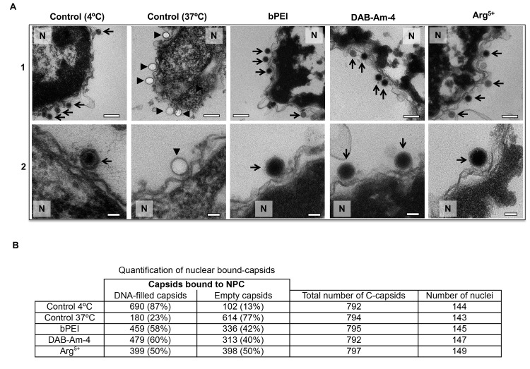
Fig 4. Ultrathin sectioning EM shows that the addition of the selected DNA condensing compounds (DAB-Am-4, bPEI 600, or Arg5+) inhibits DNA ejection from HSV-1 C-capsids into a cell nucleus through the NPC.
Positive control at 37°C shows complete DNA ejection from C-capsids in the absence of compounds (capsids were mixed with nuclei supplemented with cytosol and ATP-regenerating system). Negative control at 4°C without added compounds or ATP-regenerating system shows that no ejection occurs. In all samples, capsids and nuclei were incubated for 40 min. Bold arrows show empty capsids that ejected DNA, and thin arrows show DNA-filled capsids with DNA condensed inside. 1. Bar 500 nm. 2. Bar 90 nm. Representative EM images are shown. At least 790 capsids bound to NPCs were counted for each sample’s statistical analysis, shown in the table below.

