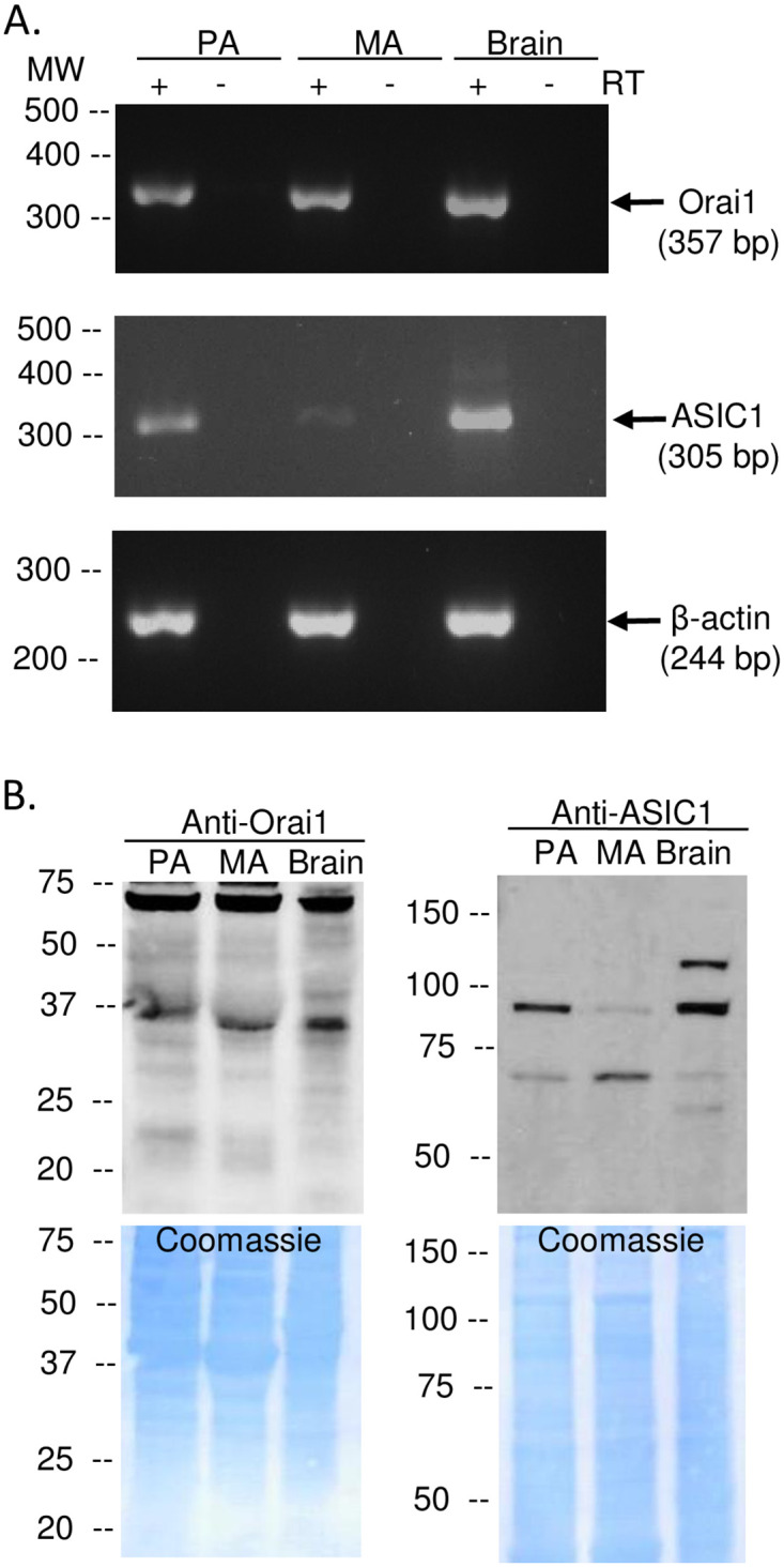
Fig 2. Orai1 and ASIC1a are expressed in pulmonary and mesenteric arteries and VSMC.

Representative PCR gels (A) showing expression of Orai1 (357 bp) and ASIC1a (305 bp) in intact pulmonary arteries (PA), mesenteric arteries (MA), and brain tissue (positive control). All lanes were loaded with 5 μL cDNA. β-actin (244 bp) was used as a loading control for intact arteries. Representative western blots (B left) showing protein expression of Orai1 and ASIC1 (~100 kDa and 60 kDa) in intact PA, MA, and brain tissue (positive control). Coomassie blue staining shows equal protein loading between samples (B; right). Each experiment was replicated 3 times.
