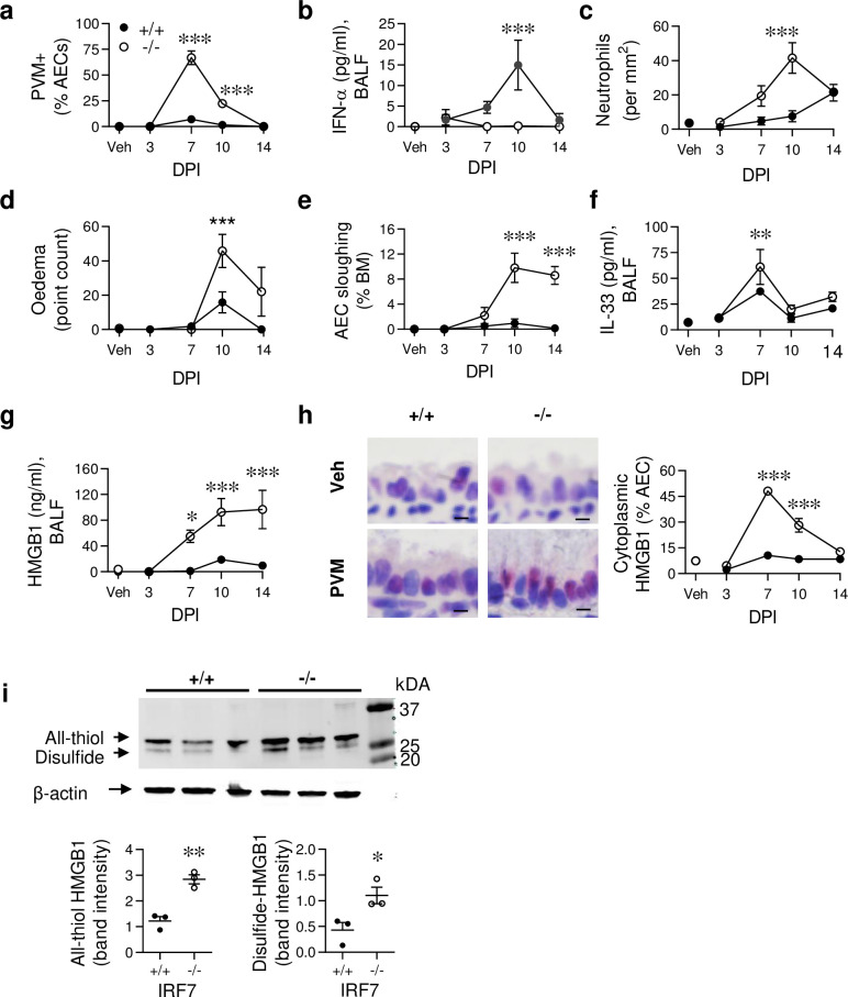
Fig 1. IRF7 deficiency impairs antiviral immunity, heightening tissue damage and alarmin release.
WT (IRF7+/+, closed circle) and IRF7-/- mice (open circle) were inoculated with PVM or vehicle at postnatal day 7 and samples collected at 3, 7, 10 and 14 days post infection (dpi). (a) Quantification of PVM+ airway epithelial cells (AECs). (b) IFN-α protein expression in BALF. (c) Ly6G+ neutrophils in lung sections. (d) Oedema. (e) AEC sloughing as a proportion of basement membrane (BM) length. (f) IL-33 and (g) HMGB1 protein expression in BALF. (h) Representative micrograph (x1000 magnification) of HMGB1immunoreactivity at 7 dpi (left panel) and quantification of cytoplasmic HMGB1 in AECs (right panel). Bars, 5 μm. (i) Electrophoretic mobility of lung homogenate loaded onto a 12% SDS-PA gel, and revealed by western blotting using polyclonal antibody against HMGB1 in PVM-infected WT (+/+) and IRF7-/- (-/-) mice at 10 dpi (top). Quantification of band intensity (bottom). Data are representative of n = 2 experiments with 4–8 mice in each group and are presented as mean ± SEM (a-g) or scatter plot (i). Data were analysed by Two-way ANOVA with Tukey post hoc test (a-g) or T-test (i); *, P < 0.05; **, P < 0.01; ***, P < 0.001 compared with the WT control group.

