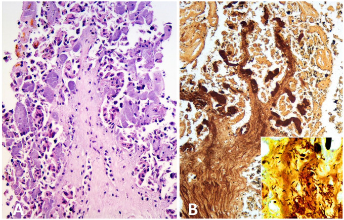
Figure 1.
Aborted ovine placenta from case D1. A. Chorionic villus with severe trophoblast necrosis, mild stromal infiltration with neutrophils, and numerous severely distended subepithelial capillaries filled with finely granular basophilic material (bacterial micro-colonies). H&E. B. Silver-stained material within severely distended subepithelial capillaries and the stroma of a necrotic chorionic villus. Inset: comma-shaped, silver-stained bacteria invading the villus stroma. Warthin–Starry silver stain.

