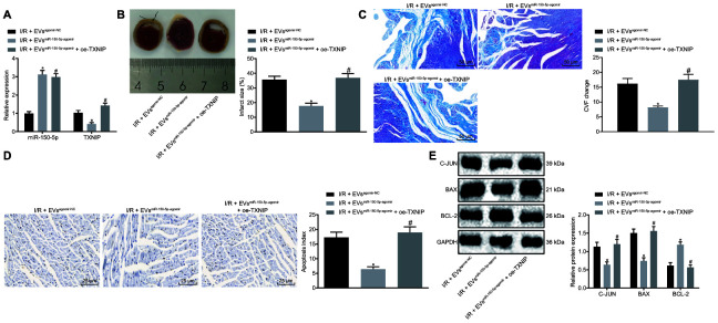
Figure 5.
The EVs-loaded with miR-150-5p/TXNIP mediates myocardial remodeling after I/R. (A) The transduction efficiency detected by RT-qPCR. (B) The myocardial infarct size detected by TTC staining. (C) The myocardial collagen detected by Masson's staining (200 ×). (D) The cardiomyocyte apoptosis detected by TUNEL staining (400 ×). (E) The protein expression of apoptosis-related factors (c-Jun, Bax and Bcl-2) normalized to GAPDH determined by Western blot analysis. * p < 0.05 vs. the I/R + EVagomir-NC group (I/R rats treated with EVagomir-NC); # p < 0.05 vs. the I/R + EVmiR-150-5p-agomir group (I/R rats treated with EV miR-150-5p-agomir). Measurement data were presented as mean ± standard deviation. Comparison among multiple groups was analyzed by one-way analysis of variance, followed by Tukey’s post hoc test. n = 12.

