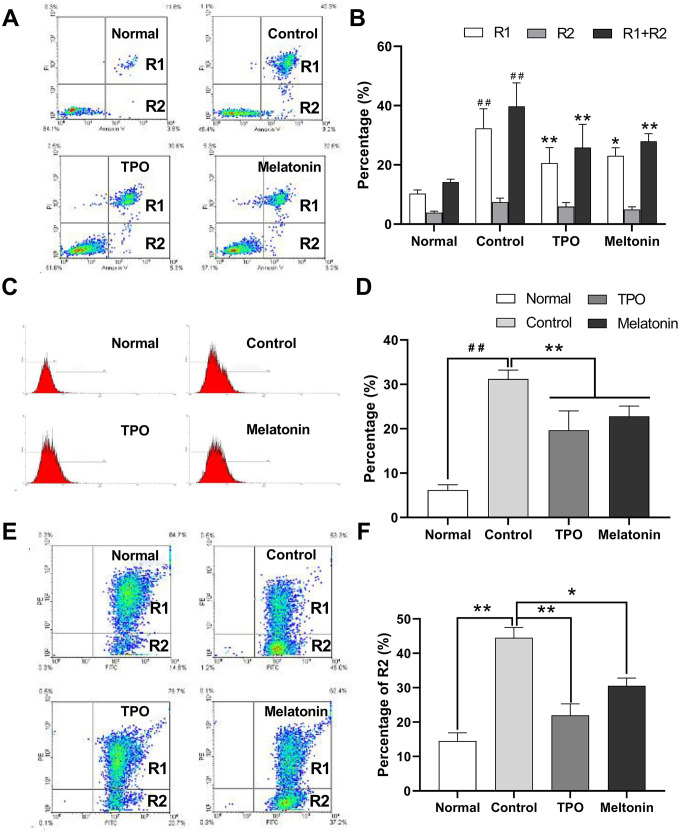
Figure 2.
Melatonin significantly suppresses the apoptosis of nutrition-depleted CHRF cells. Cells were treated with saline (control), melatonin (200 nM) or TPO (positive control, 100 ng/mL) for 72 h. CHRF cells were harvested and subject to assays to measure the levels of (A) Annexin/PI (n=4), (C) activation of Caspase-3 (n=3) or (E) polarized mitochondrial membrane potential (JC-1, n=3). (B), (D, F) were the statistical analyses of (A, C, E), separately. Normal, CHRF cells incubated with 10% FBS; Control, CHRF cells incubated with 0.5% FBS. One-way ANOVA or Two-way ANOVA (with a Tukey multiple comparison test) was employed to test for significance. # # Compared with normal group, p< 0.01; * p < 0.05, ** p < 0.01.

