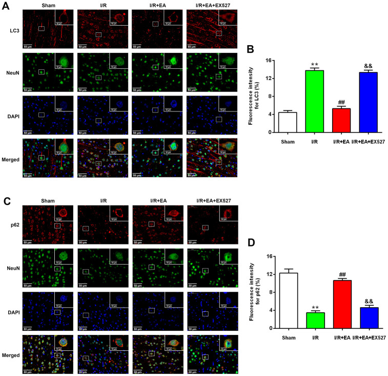
Figure 4.
EA pretreatment inhibited the expression of LC3 in NeuN-positive neurons of the peri-ischemic cortex, while promoted the expression of p62 in NeuN-positive neurons of the peri-ischemic cortex after 24 h of reperfusion. (A) Representative images of LC3 (red)/NeuN (green) double-labeled staining (400×). High magnification images are shown in the small windows (1000×). Scale bar, 400×: 50 μm; 1000×: 10 μm. (B) Mean fluorescence intensity for LC3 in neurons of the peri-ischemic cortex. (C) Representative images of p62 (red)/NeuN (green) double-labeled staining (400×). High magnification images are shown in the small windows (1000×). Scale bar, 400×: 50 μm; 1000×: 10 μm. (D) Mean fluorescence intensity for p62 in neurons of the peri-ischemic cortex. Data were presented as the mean ±SEM (n=3). **P<0.01 vs. sham group. ##P<0.01 vs. I/R group; &&P<0.01 vs. I/R + EA group.

