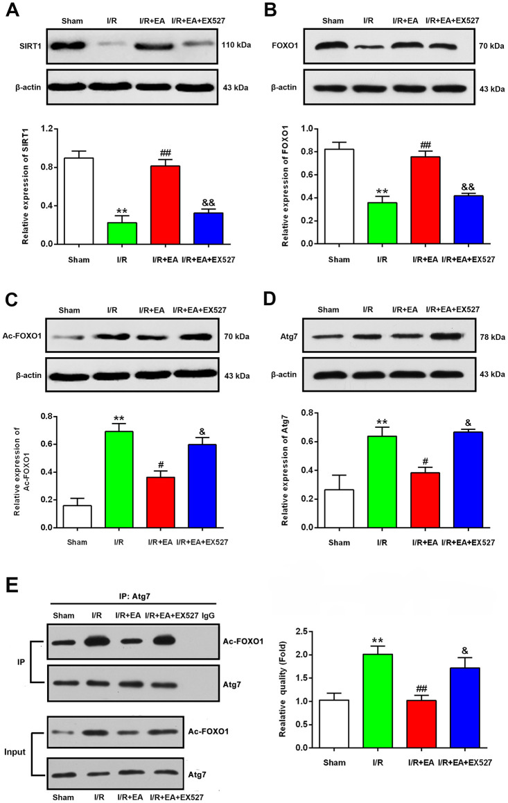
Figure 6.
EA pretreatment up-regulated the protein levels of SIRT1, FOXO1, down-regulated the protein levels of Ac-FOXO1 and Atg7 in the peri-ischemic cortex after 24 h of reperfusion. (A) Protein band and relative expression of SIRT1 in the peri-ischemic cortex. (B) Protein band and relative expression of FOXO1 in the peri-ischemic cortex. (C) Protein band and relative expression of Ac-FOXO1 in the peri-ischemic cortex. (D) Protein band and relative expression of Atg7 in the peri-ischemic cortex. (E) Co-IP between Atg7 and Ac-FOXO1 in the peri-ischemic cortex and the relative quality of Ac-FOXO1 after normalization with Atg7, and input results of Atg7 and Ac-FOXO1. β-actin was used as a loading control. Data were presented as the mean ± SEM (n=3). **P<0.01 vs. sham group. #P<0.05 and ##P<0.01 vs. I/R group; &P<0.05 and &&P<0.01 vs. I/R + EA group.

