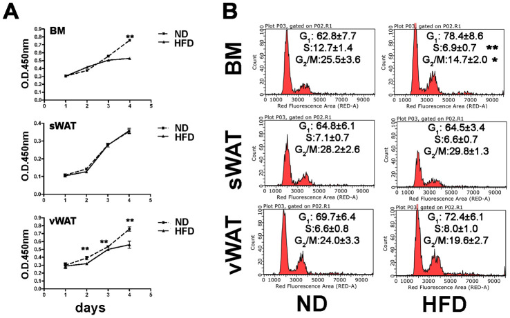
Figure 2.
Proliferation and cell cycle analyses. (A) MSC cell proliferation was evaluated by Cell Counting Kit-8 (CCK-8) colorimetric assay. The graph shows data coming from obese and control samples. Data are shown with standard deviation (SD) n=6 animals for each experimental condition, **p<0.01. (B) Representative cell cycle analysis of MSCs harvested from obese and normal mice. Data are expressed with SD (n=6 animals for each experimental condition) *p<0.05, **p<0.01.

