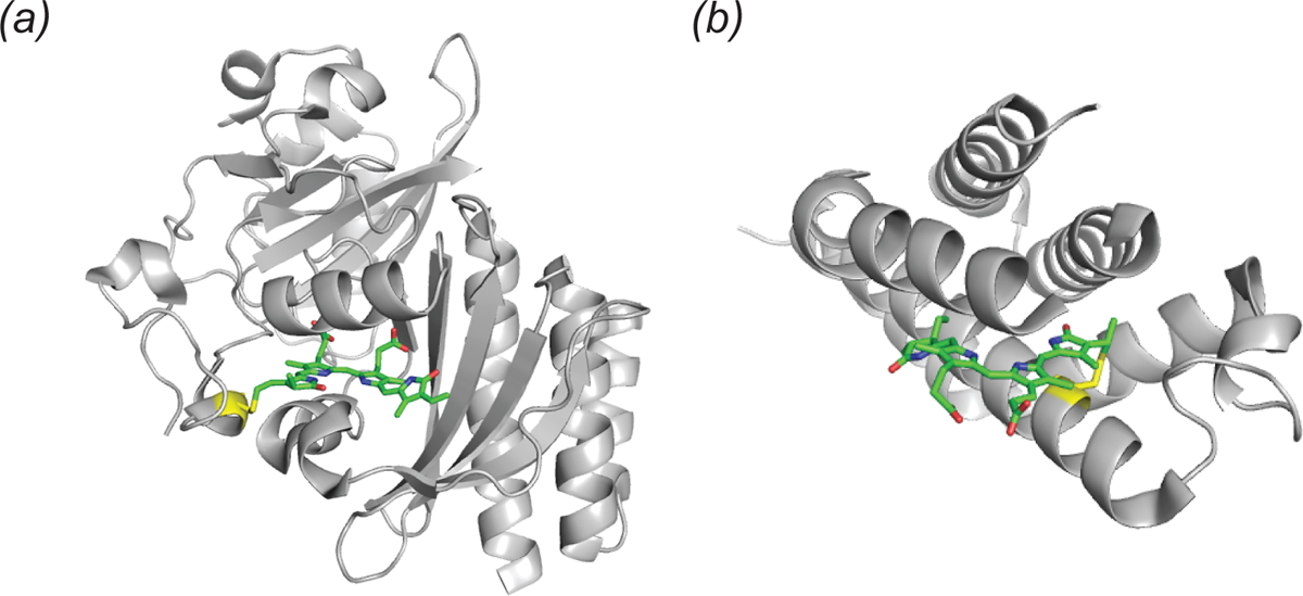
Fig. 3.

Structure of two fluorescence activating proteins bound to biliverdin. (a) Cartoon representation of the infrared fluorescent protein IFP 2.0 (PDB ID: 4CQH). The biliverdin chromophore (green sticks) is shown with the covalent linkage to cystine 24 (yellow). (b) Cartoon representation of smURFP (PDB ID: 6FZN). The biliverdin chromophore (green sticks) is shown with the covalent linkage to cystine 52 (yellow sticks).
