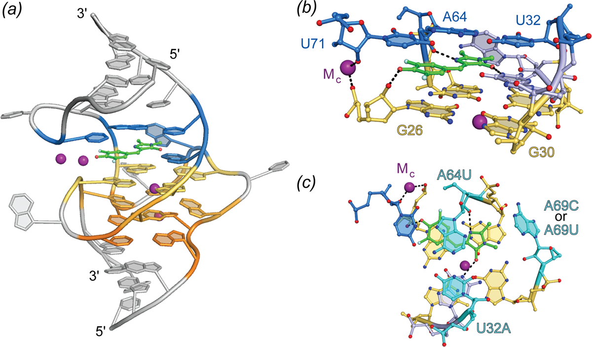
Fig. 5.

Structure of Spinach aptamer. (a) Cartoon representation of the Spinach aptamer core (PBD ID: 4TSO). The DFHBI fluorescent ligand is shown as green ball-and-stick with flanking A-form helices and unstructured nucleotides (grey), G-quadruplex nucleotides (warm colors), triplex and flanking binding pocket nucleotides (blue), shown in cartoon representation. Bound K+ ions represented as purple spheres. (b) Side view of the Spinach binding pocket (ball-and-stick) colored according to (a). Hydrogen bonds and metal coordination are represented as black dashed lines. (c) Top view of the Spinach ligand-binding pocket. Residues demonstrated to alter spectral properties in HBI-derivative binding aptamers are colored cyan and labeled with mutation details.
