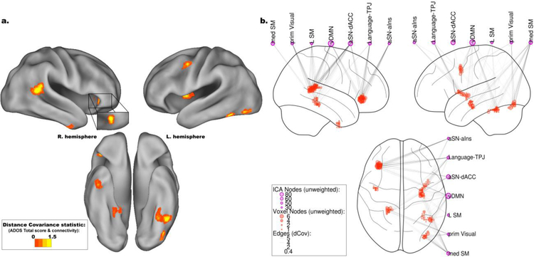
Figure 2.
Neuroanatomical and network associations between autism spectrum disorder symptoms and functional connectivity as measured by distance covariance (dCov). a. Throughout the entire brain, Autism Diagnostic Observation Schedule (ADOS) total score was primarily associated with connectivity to the right temporoparietal junction (TPJ) and anterior insula (aIns), as well as the left fusiform gyrus, middle frontal gyrus, and middle insula. Smaller clusters of association were in the right temporal pole and lingual gyrus, as well as left posterior insula and lateral occipital lobe.
b. Seven Intrinsic Connectivity Networks (ICNs), identified with Independent Components Analysis (ICA), primarily contributed to the neuroanatomical associations displayed in Figure 2a. These included medial and Left Sensorimotor networks (med SM and L SM, respectively), primary Visual (prim Visual), ventral Default Mode Network (vDMN), a Language subnetwork centered on the bilateral temporoparietal junction (Language-TPJ), and two subnetworks of the anterior Salience Network (aSN) centered on the dorsal Anterior Cingulate Cortex (aSN-dACC) and anterior insula (aSN-aIns). The influence of connectivity on these ICNs varied by region. The left fusiform gyrus was associated with ADOS total scores through its connections to prim Visual and med SM networks, while the aIns was associated with ADOS total scores through both aSN subnetworks, the vDMN, and the Language-TPJ network.

