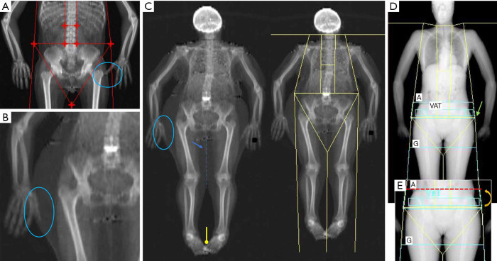
Figure 2.
Examples of common positioning and postprocessing artifacts that may limit the accuracy of whole body DXA scan. (A,B) Two examples of superimposition between fingers and leg fat tissue (see the blue circles). (C) Overweight patients in which positioning may result complicated: in this case there is an overall superimposition between several anatomic regions such as fingers with subcutaneous fat (blue circle), foots (yellow line) and legs (blue arrow). The latter may create problems in separating the fat mass content between the two legs (dashed blue line). (D) Inaccurate placement of pelvis line (green arrow), which is located just above both femoral heads; this create problems in the correct identification of android and gynoid regions, which is typically done automatically by the software. (E) Image shows the correct placement of the pelvic line (dashed red line), after image correction (indicated by curved arrow).

