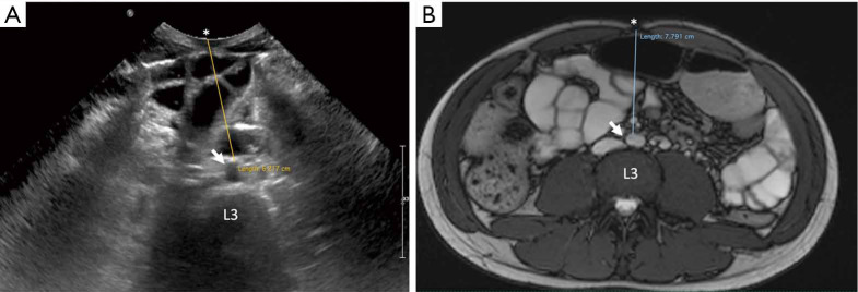
Figure 3.
Ultrasound (A) and abdominal-MRI (B) of a 15-year-old boy. After treatment follow-up of Crohn’s disease (same patient as in Figure 2). (A) The VAT thickness is measured on the transverse plane at the level of the umbilicus (linea alba) (*) as the distance from the umbilicus to the anterior wall of the aorta (white arrow) at the level of the L3 vertebral body. (B) Correlation with the MRI image at the same level of the ultrasound image with the MRI measurement on true fast imaging with steady state free precession (True-FISP) image. Note the variation in magnitude of the fat measurement in the same patient using the two different techniques (even though this was performed during the same diagnostic session). Ultrasound is an operator-dependent technique, and as such, technique has to be precise and standardized (supine position, expiration, arms along the body), otherwise error may be introduced. VAT, visceral adipose tissue.

