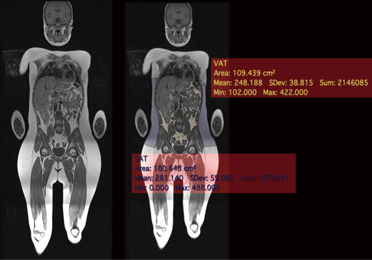
Figure 5.
Whole-body MRI in a 12-year-old girl (unremarkable examination performed to rule out bone marrow lesions with the suspicion of a Chronic Recurrent Multifocal Osteomyelitis). Single axial T1-weighted coronal whole-body image was post-processed with the semi-automatic free resource software Horos (https://horosproject.org) using the “brushing tool”. VAT can be distinguished from SAT. The values are expressed in cm2. The region of interest for the measurements is the android region, defined as a segment of the abdomen comprised between a lower demarcation line joining the superior limits of the iliac crests and an upper demarcation line drawn at 20% of the distance in between the iliac crests line and the mentum (chin). VAT, visceral adipose tissue; SAT, subcutaneous adipose tissue.

