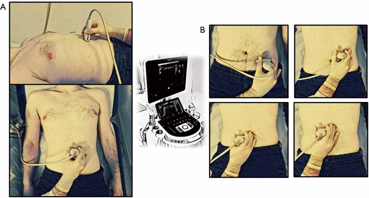
Figure 2.
On the left (A) is represented the anatomical posture of ultrasound’s probe in the evaluation of IAFT, while on the right (B) is represent represented the anatomical postures of ultrasound’s probe in the evaluation of MFT. IAFT, intra-abdominal fat thickness; MFT, mesenteric fat thickness.

