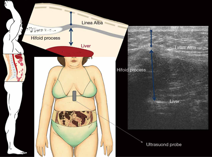
Figure 3.
On the left are represented anatomic pictures of the abdominal sagittal section (with its enlargement in upper) and of the abdominal frontal section where MinASFT (blue caliper with diamond tips) and maximum preperitoneal fat thickness (blue caliper with arrowheads) have been measured with US. US, ultrasonography; MinASFT, minimum abdominal subcutaneous fat thickness.

