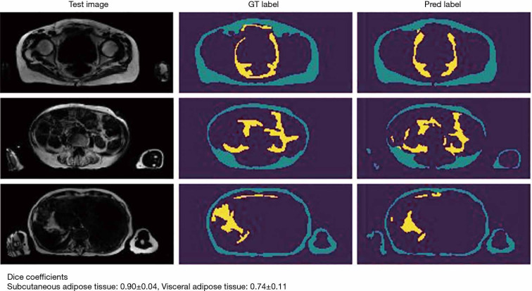
Figure 2.
Exemplary slices of a segmented whole-body MRI of a healthy 50-year-old female, acquired as axial T1-weighted gradient-echo Dixon sequence. The first column shows the original slices, second and third columns are color-labeled maps of the segmentation masks. Adipose tissue was segmented in an experimental approach, differentiating between subcutaneous fat (green) and visceral adipose tissue (yellow). The second and third columns from the left represent manual ground truth (GT) and predicted automatic (Pred) label mask. The comparability was excellent between GT and Pred label using Dice coefficient, however, the agreement was better for subcutaneous fat (0.90±0.04), compared to visceral fat (0.74±0.11).

