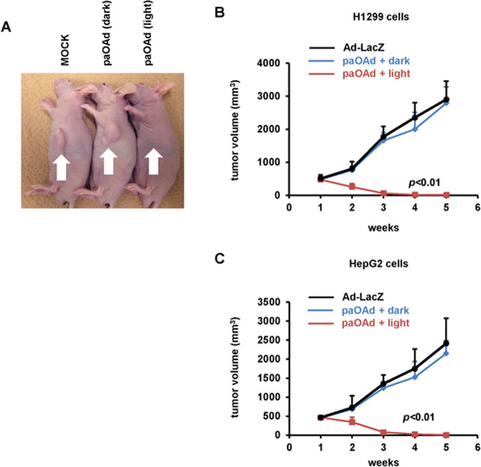
Fig. 2. In vivo antitumor effects of paOAd in a subcutaneous xenograft model mice.
BALB/c nu/nu mice bearing H1299 or HepG2 subcutaneous xenograft tumors were intratumorally administered 107 IFU of paOAd at days 0 and 3. H1299 xenograft tumors were irradiated with 90 mW/cm2 blue light from day 0 to day 10 (8 h/day). a The macroscopic appearance of H1299 tumors in nu/nu mice at 5 weeks after treatment. b, c Growth of H1299 (b) or HepG2 (c) xenograft tumors was expressed as the mean tumor volume ± S.E. Data were generated from 16 mice for each group. Replication-deficient LacZ-expressing Ad (Ad-LacZ) was used as a control. Statistical significance was evaluated by two-way repeated ANOVA followed by Tukey’s post hoc tests. At 2, 3, 4, and 5 weeks after the illumination, the mean tumor volumes in the “paOAd + light” group were higher than those of the other groups (p < 0.01).

