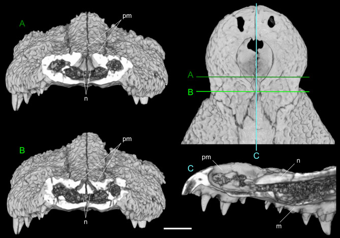
Figure 6.
CT investigation of the relationship between the nasals and the premaxillae in sn813/lj. Top left: anterior tip of the snout in dorsal view showing the planes of the sections in (A), (B) and (C). (A) anterior transversal section do not involve the nasals; (B): posterior transversal section cutting the nasals and showing that the premaxillae are entirely separated by the nasals; (C): sagittal section showing that the nasals are slightly lowered in the area of the posterior process of the premaxillae. Note in (A) and (B) how the nasals are raised to form the medial boss of the snout. Scale bar: 3 cm.

