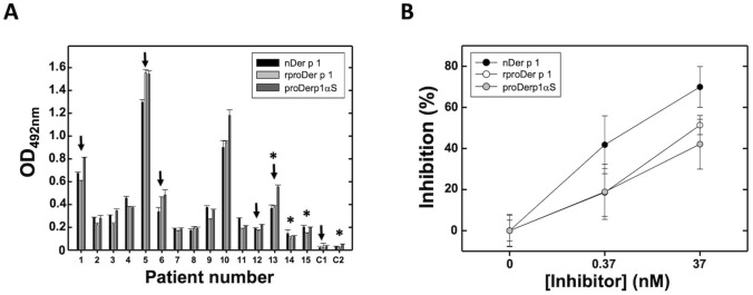
Figure 3.
Human serum IgE recognition assays. (A) nDer p 1, rproDer p 1 and proDerp1αS comparative detection by serum IgE of Der p 1-positive allergic patients (#1–15) and non-allergic (C1, C2) individuals by mean of indirect ELISA. Black arrows indicate selected sera for in vitro humRBL-2H3 degranulation and viability assays. Asterisks indicate selected patients for BAT and basophils cytotoxicity assays. (B) Inhibition of serum binding to nDer p 1 by rproDer p 1 (open circles), the derived immunotoxin chimera (grey circles) and using the allergen nDer p 1(black circles) as a control of maximum inhibition. Graph representation shows the resulting media ± standard deviation for three sera (#1, 3, 10). All experiments were done by duplicate.

