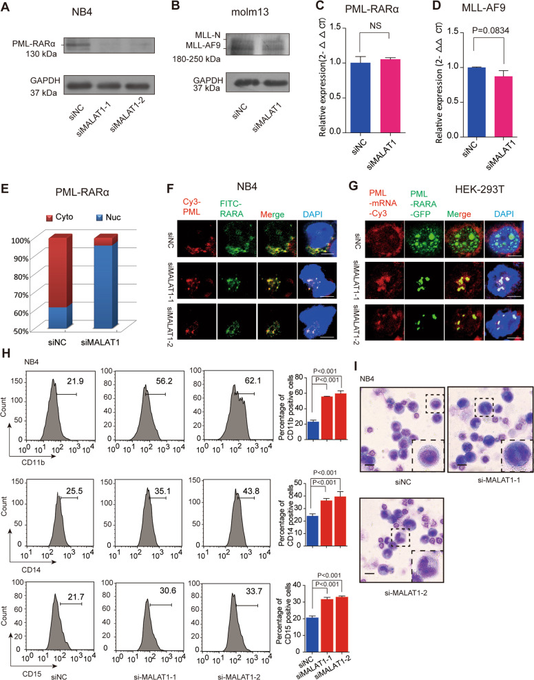
Fig. 2. Knockdown of MALAT1 significantly repressed the expression levels of fusion proteins by promoting their mRNAs retention in nuclei.
a, b Protein levels of PML-RARα and MLL-AF9 were reduced after knockdown of MALAT1 in NB4 cells and Molm13 cells, respectively. GAPDH was used as a loading reference. All experiments were performed independently at least three times. c, d qRT-PCR showed the mRNA levels of PML-RARα and MLL-AF9 were not markedly changed after reducing MALAT1 levels via siRNA. Data are shown as the means ± s.e.m.; n = 3 independent experiments. e PML-RARα mRNA detection by qRT-PCR after separating the nuclear and cytoplasmic RNAs. f PML-RARα mRNA location detecting by RNA FISH in NB4 cells. Two different fluorophores were added to PML (Cy3, red) and RARα (FITC, green) mRNA, respectively. The dots merged into yellow dots represent the mRNAs of PML-RARα. Scale bar represents 4 μm. g RNA FISH experiments were carried out according to the process shown in Fig. 1b in HEK-293T cells. Scale bar represents 4 μm. h Cellular differentiation markers CD11b, CD14, and CD15 were detected by flow cytometry. The results showed that compared to the negative controls, both markers were increased significantly when MALAT1 was down-regulated. The data were analyzed from at least three independent experiments and are shown as the means ± s.e.m. i Wright–Giemsa staining showed that cellular differentiation was significantly promoted after reducing MALAT1 levels in NB4 cells. Scale bar represents 20 μm.

