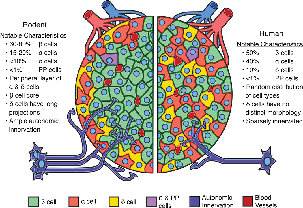
Figure 1. Comparative architecture of pancreatic islets of mice and humans.
Pancreatic islets of mice and humans differ in important ways, but also share many features in common. These shared features make mouse islets useful experimental models to study many aspects of human islet biology. The relative proportions of endocrine cell types in mouse (left) and human islets (right) are similar with beta cells (β; green) comprising the majority of the islet cell mass followed by alpha (α; light red) and delta cells (δ; yellow). Other islet endocrine cells such as pancreatic polypeptide and epsilon cells (PP and ε; purple) are more sparse in number. Human islets occur in a wide variety of sizes and conformations that range from highly structured to more random distributions of cells. Mouse islets exhibit a more uniform architecture with alpha and delta cells at the islet periphery surrounding a beta cell core. Islets in both species are vascularised (dark red) and innervated (dark blue) for rapid sensing of changing energy needs, although mouse islets are more densely innervated than humans.

