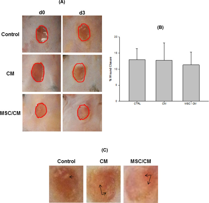
Figure 4. Evaluation of wound closure after MSC transplantation.
Wounds were evaluated before (d0) and after (d3) MSC transplantation. Wound closure was compared between the same experimental group (circle of the same size) (A). Wound closure was determined by using the ImageJ program. It was expressed as the percentage (means ± SE) of wound closure at day 3 as compared with d0 in each group (% of wound closure = [wound area d0 − wound area d3/area d0] × 100). There were not a statistically significant difference in wound closure between day 3 and d0 post-wounding in all groups (control, n=5; CM, n=5 and MSC/CM, n=4) (B). Signs of early re-epithelialization (whitish areas covering the wound surface) were observed in wounds (higher magnification) in all groups (C, arrows).

