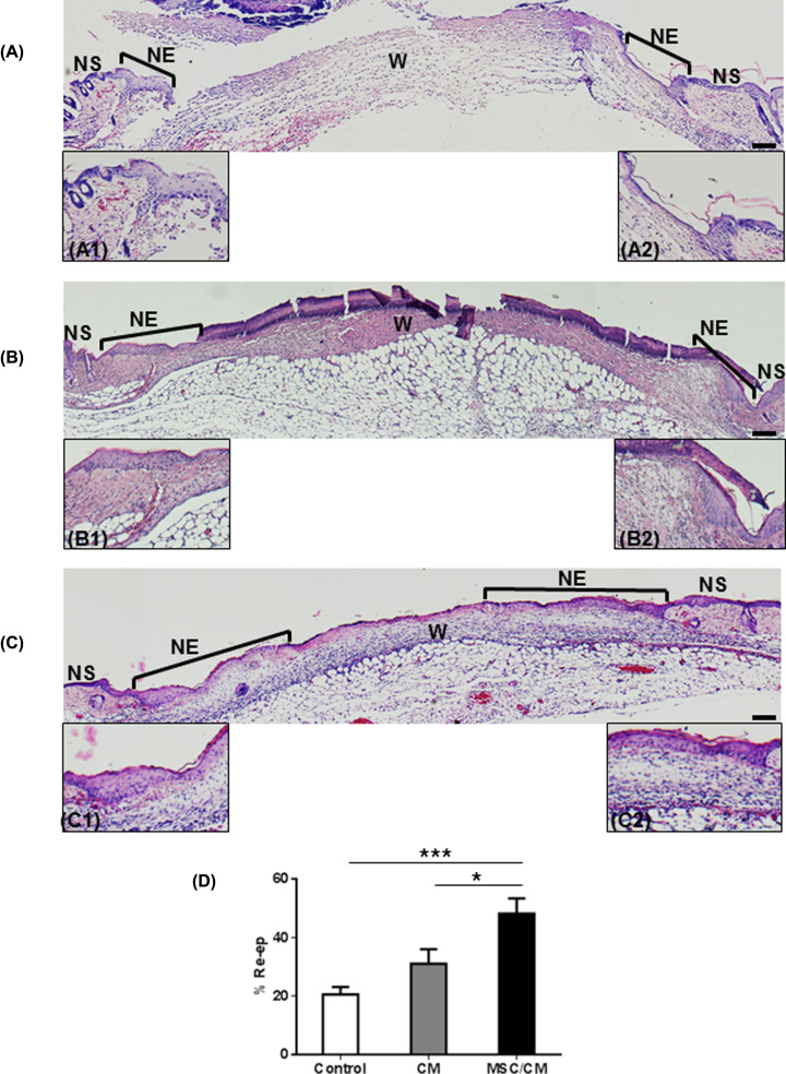
Figure 5. MSC transplantation enhances wound re-epithelialization.
Histological studies of wounds were performed in untreated wounds (control, A), CM-treated wounds (B) and MSC/CM-treated wounds (C), at day 3 post wounding. H&E-stained sections show NS and NE in the edges of wounds (W). Higher magnification of the newly formed epidermis is shown in each section (control, A1–A2; CM, B1–B2; and MSC/CM C1–C2). Histologic sections of wounds treated with MSC/CM show a larger area of re-epithelialization (C), as compared with those treated with CM alone (B) or control (A). Image analysis from histological sections show a significant increase in the percentage of re-epithelialization in wound treated with MSC/CM, as compared with control groups (D). Scale bar = 100 μm. Results are presented as means ± SE (Control, n=7; CM, n=7; MSC/CM, n=8). *P<0.05; ***P<0.001.

