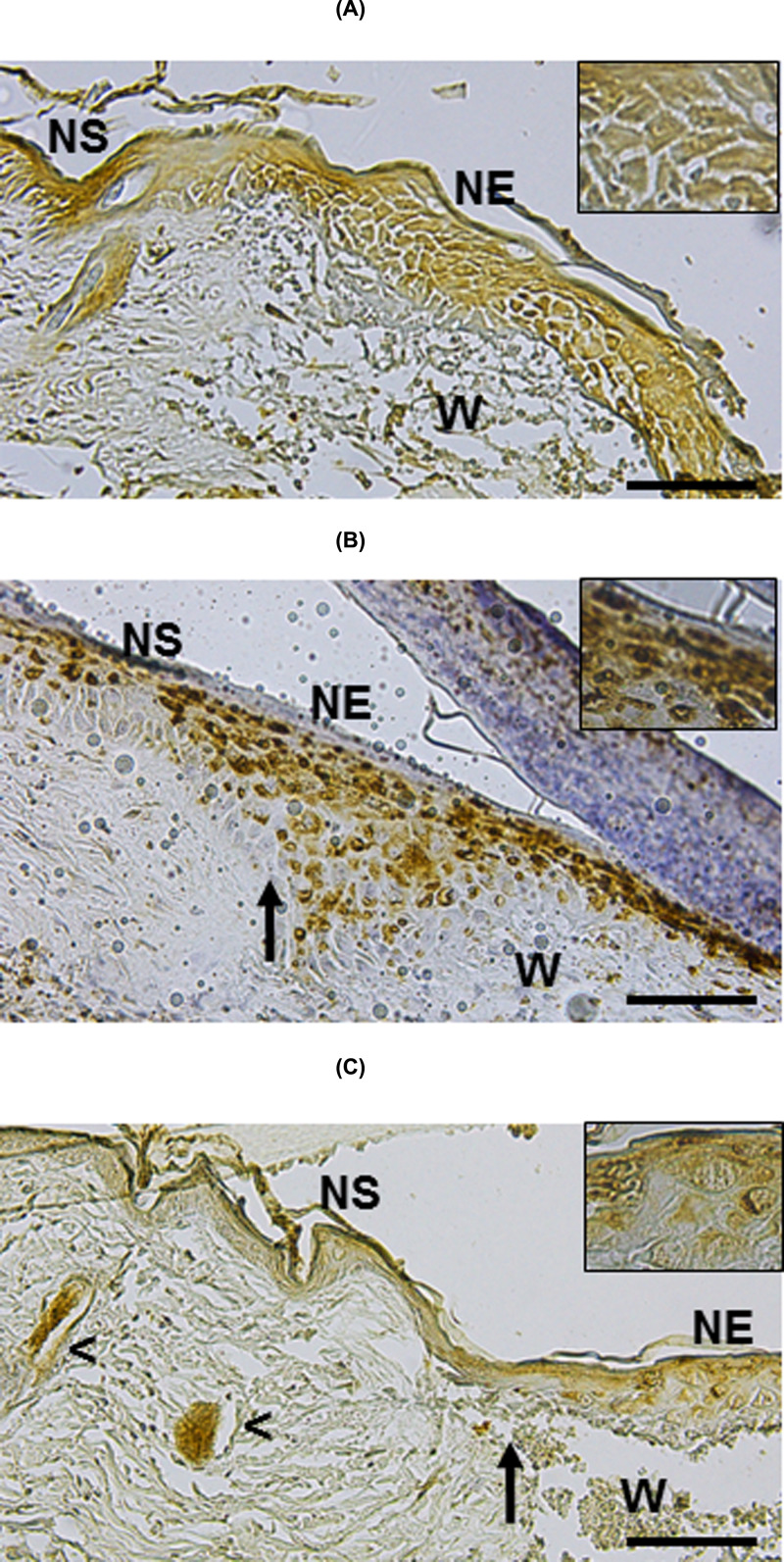
Figure 7. Detection of Lgr6+ progenitor cells in cutaneous wounds after 3 days of MSC implantation.
IH studies to detect Lgr6+ cells were performed in untreated wounds (control, A), CM-treated wounds (B) and MSC/CM-treated wounds (C). Tissue sections show NS and NE in the edges of wounds (W). Lgr6+ cells were present at the NS and NE of the control group and treated with CM (A,B, respectively). Most of the Lgr6+ cells were detected at the NE adjacent to the wound treated with MSC/CM (C). They were also detected in HFs close to the wound (head arrows). Each picture is representative of three different experiments, all with similar results. Scale bar = 50 μm.

