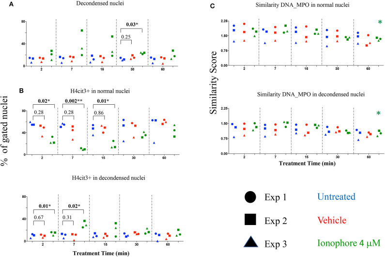
Figure 3.
Calcium ionophore treatment induces a rapid change in histone citrullination in both normal and decondensed neutrophil nuclei in the absence of extracellular calcium. Neutrophils purified from three healthy donors were left in DPBS (blue), or treated for the indicated times with vehicle only, DMSO (red), or A23187 4 μM (green) (A) Percent neutrophils with decondensed nuclei. (B) Percent neutrophils with normal and decondensed nuclei positive for H4cit3. (C) Similarity scores for DNA (Hoechst stain) and MPO, marking co-localization of two distinct cellular compartments, nucleus, and type I granules. Paired t-test was used to compare untreated with vehicle and ionophore 4 μM, respectively at each time point; Anova for panel ionophore-mediated response in (C) *P < 0.05, **P < 0.01.

