
Fig. 1.
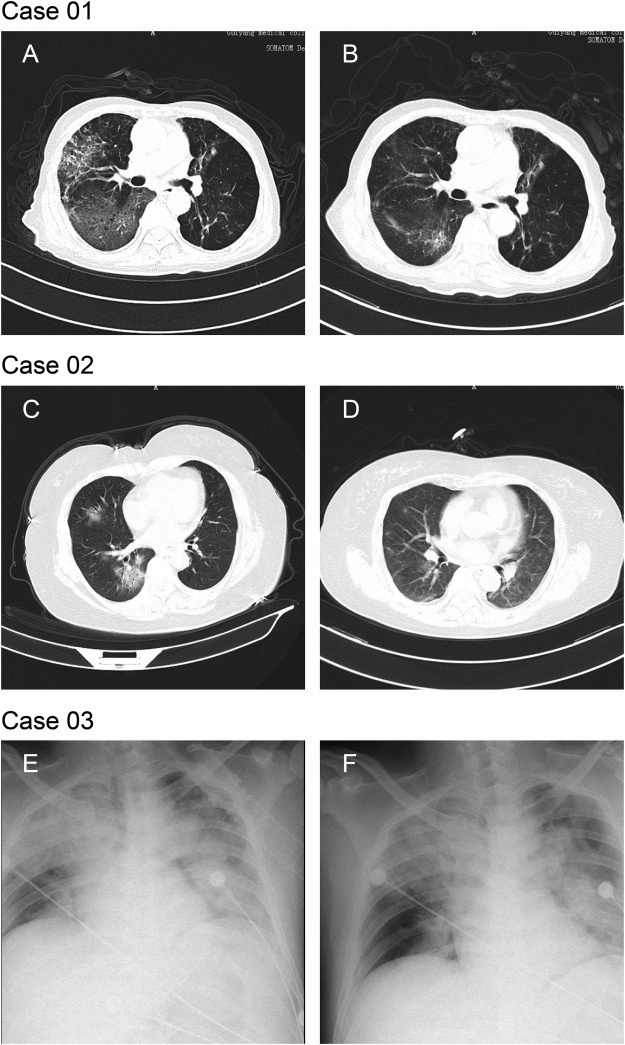
Chest CTs and x-rays of three patients
Case 01: Chest CTs was obtained on 2nd and 14th day post admission. CT findings of Case 01 obtained on 2nd day post admission showed multiple mottling and groundglass opacities (GGO) in bilateral lungs, predominantly involving right lobes. Septal thickening and extensive consolidation in the right middle and lower lobes presented a “paving stone like” reticulation (1A). Chest CT images for 14th day post admission showed improved status with bilateral ground-glass opacity, whereas partial consolidation had been resolved (1B).
Case 02: Chest CTs was obtained on 2nd and 14th day post admission. CT scan of Case 02 obtained on 2nd day post admission showed mixed multifocus GGO and consolidation in basal segment of right lower lobe, and GGO with minimal reticulation in right middle lobe (1C). CT scan for 14th day post admission showed healing of the consolidation and GGO, remaining linear opacities (1D).
Case 03: Chest x-rays was obtained on the first day post admission. The brightness of both lungs was diffusely decreased and extensive patchy shadows were observed, edges were blurred and the heart shadow enlarged slightly. Right diaphragmatic surface was light and smooth, costal diaphragmatic angle was sharp, while left diaphragmatic surface and costal diaphragmatic angle blunted (1E and 1F).