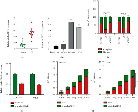
Figure 1.

High expression of circFAT1(e2) was detected in OS tissues and cells, and circFAT1(e2) promoted cell proliferation. (a) qRT-PCR analysis for circFAT1(e2) expression in 10 pairs of osteosarcoma and normal tissue. (b) CircFAT1(e2) expression was higher in osteosarcoma cells than in normal cell lines (hFOB 1.19). (c) CircFAT1(e2) was mainly distributed in the cytoplasm of OS cells. (d) The expression of circFAT1(e2) in osteosarcoma cells transfected with si-circFAT1(e2) decreased by nearly half. (e) The CCK-8 assay showed that the knockdown of circFAT1(e2) reduced the proliferation rate of OS cells. Error bars represent the mean ± SD of at least three independent experiments. ∗p < 0.05, ∗∗p < 0.01 vs. control group.
