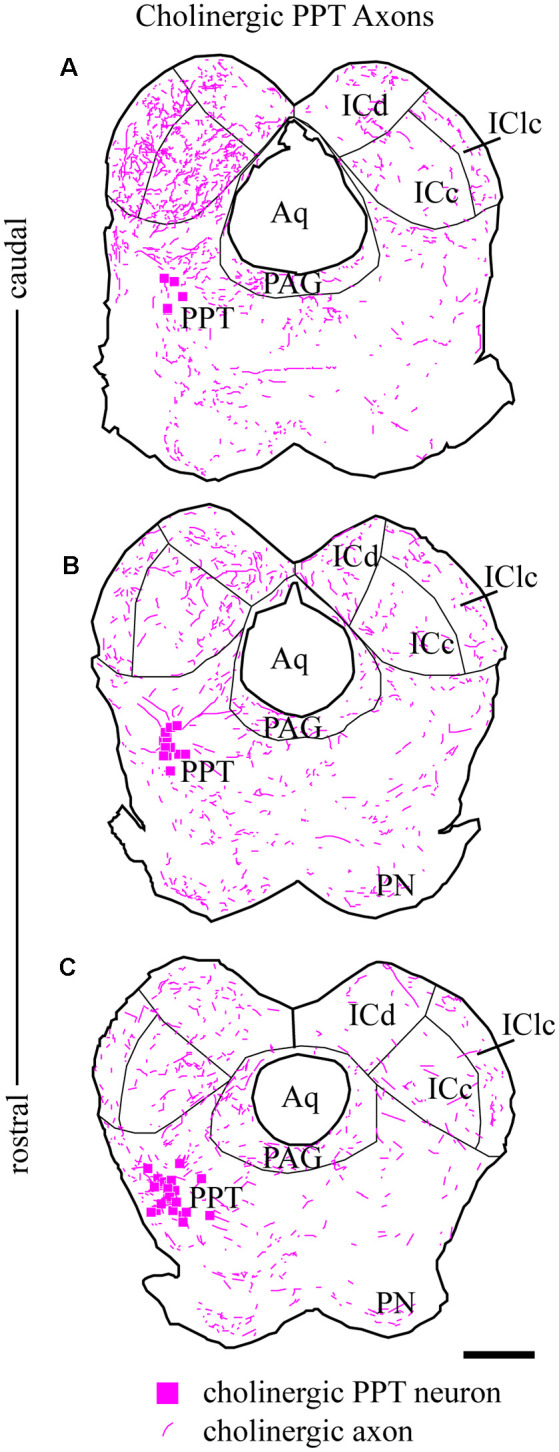
Figure 3.

Cholinergic axons travel through the tegmentum to reach ipsilateral and contralateral IC. Fluorescent-labeled axons (magenta lines) were observed throughout transverse IC sections after labeling cholinergic cells in the left PPT (magenta squares). The plots show the axons in a series of transverse sections through the IC at caudal (A), middle (B), and rostral levels (C). On both sides, labeled axons appear to enter the IC all along its ventral border. Sections are 40 μm thick and 240 μm apart. Scale bar = 1 mm.
