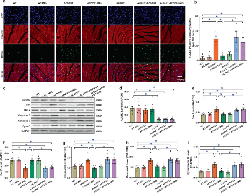
Fig. 6.
Effect of melatonin on apoptosis levels in WT, APP/PS1, ALDH2-knockout (ALDH2−/−), and ALDH2−/−-APP/PS1 mice. a Representative image of TUNEL; b TUNEL-positive cardiomyocytes per 100 cells; c representative gel depicting ALDH2, the apoptosis markers Bax, Bcl-2, Caspase 3, Caspase 9, and cytochrome C and GAPDH (loading control) levels using specific antibodies; d ALDH2 expression; e Bax level; f Bcl-2 level; g Caspase 3 level; h Caspase 9 level; and i cytochrome C level. The data are shown as the mean ± SEM, n = 5–8 mice per group. *p < 0.05 between the indicated groups

