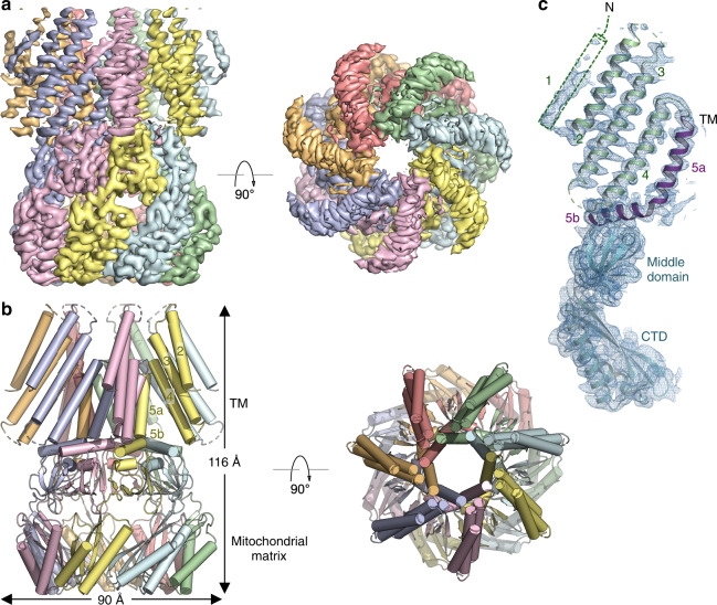
Fig. 1. Cryo-EM structure of AtMSL1.
a Orthogonal views of the cryo-EM density contoured at 7.0 σ. Each subunit is uniquely colored. b Orthogonal views of the MSL1 structure. Transmembrane helices TM2-5, the C-terminal extramembrane domain located in the mitochondrial matrix, and the dimension of the channel are indicated. c A single subunit of MSL1 is shown in cartoon representation with TM helices (TM2-4 in green and TM5 in purple) and extramembrane domains (cyan) indicated. Cryo-EM density contoured at 6.5 σ is shown in blue mesh.

