
Figure 3.
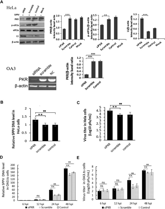
PKR knockdown increases SPPV replication. (A) HeLa cells were transfected with the siRNA specific for PKR (75 pmol/mL) or control siRNA. Take HeLa cells transfected with Lipo3000 as control. After 48 h of incubation, cells were infected with SPPV Gulang/2009. Take uninfected cells as MOCK. PKR, phosphorylated eIF2α, and SPPV L1 expression were detected by western blotting. β‐Actin was used as a protein loading control. (B) SPPV genome DNA levels in the cells were quantified by real‐time PCR at 12 hpi, which were transfected with siPKR or control siRNA and then infected with SPPV at 48 hpt. HeLa cells were transfected with Lipo3000 as control. (C) HeLa cells were transfected with siPKR or control siRNA and infected with SPPV Gulang/2009 at 48 hpt. (D) SPPV genome DNA levels in the sheep OA3 were quantified by real‐time PCR at 6, 12, 24, and 48 hpi, which were transfected with siPKR or control siRNA and then infected with SPPV at 48 hpt. OA3 were transfected with Lipo3000 as control. (E) OA3 cells were transfected with siPKR or control siRNA, infected with SPPV Gulang/2009 at an MOI of 3 at 48 hpt. Cells and virus were collected at 6, 12, 24, and 48 hpi. SPPV titer in cells was assayed by plaque assay. Data are represented as mean ± SEM; n = 3. Representative of three independent experiments. Significance was analyzed using two‐tailed Student's t‐test. **P < 0.01; ***P < 0.001. ns, not significant.