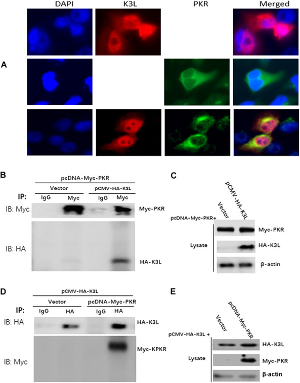
Figure 6.

K3L interaction with PKR. (A) The colocalization of PKR and K3L in HeLa cells. HeLa cells was transfected with pCMV‐SPPV K3L plasmids (2 μg, upper panel) and pcDNA‐sPKR plasmid (1 μg, middle panel) for 36 h, respectively. HeLa cells were cotransfected with pCMV‐SPPV K3L plasmids (2 μg) and pcDNA‐sPKR plasmid (1 μg) for 36 h (lower panel). Expression of pCMV‐SPPV K3L and pcDNA‐sPKR was detected by immunofluorescence assay. Cells were double‐immunostained for pCMV‐HA‐SPPV K3L (red) and pcDNA‐sPKR (green); the cellular nuclei were counterstained with DAPI (blue). (B) HeLa cells were cotransfected with pcDNA‐Myc‐sPKR and pCMV‐HA‐K3L plasmids for 36 hours. Whole cell lysates were immune‐precipitated with mouse normal IgG antibody or mouse anti‐Myc antibody and subjected to western blotting. (C) Whole cell lysates were also detected with western blotting to validate the expression of the proteins of interesting. (D) HeLa cells were cotransfected with pcDNA‐Myc‐sPKR and pCMV‐HA‐K3L plasmids for 36 hours. Whole cell lysates were immune‐precipitated with mouse normal IgG antibody or mouse anti‐HA antibody and subjected to western blotting. (E) Whole cell lysates were also detected with western blotting to validate the expression of the proteins of interesting.
