
Figure 1.
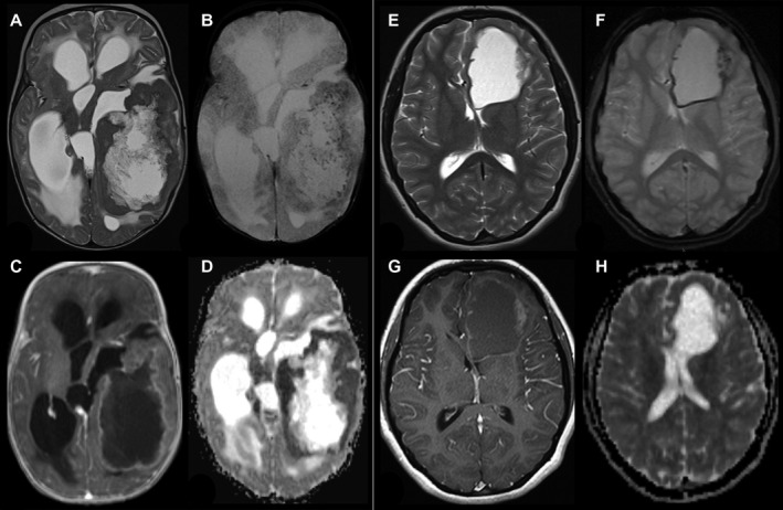
Radiological findings in two patients with YAP1‐MAMLD1‐fused ependymoma. (A–D; Patient #9) 4 months old infant with a huge left temporal tumor, a large cystic part surrounded by solid tumor, which is isointense to gray matter on T2‐weigthed image (A) and punctuated hemorrhages in the solid part a small rim along the cyst wall (T2*, B). The solid part of the tumor shows a garland‐shaped enhancement (C) and ADC values are decreased compared to gray matter. T2 and ADC changes are compatible with a higher cell density of the tumor. (E–H, patient # 6) 15‐year‐old girl with a left frontal tumor without edema consisting of a large cyst and a small solid part at the lateral border. The solid part is nearly isointense to gray matter in T2‐weighted image (E). T2* (F) reveals subtle punctuated hemorrhages in the solid part a small rim along the cyst wall. The solid part of the tumor shows a faint enhancement on T1‐weighted image (G) and the apparent diffusion coefficient (ADC) is comparable to the gray matter.