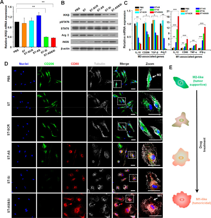
Figure 4.
Repolarization of M2-like macrophages in vitro. (A) IKKβ mRNA levels determined by quantitative real-time PCR (qRT-PCR) in M2-like macrophages incubated with different formulations for 48 h. **P < 0.01. (B) Protein expression levels of IKKβ, STAT6, Arg I, and iNOS determined by Western blot (WB) in M2-like macrophages incubated with different formulations for 48 h. (C) mRNA levels of M2/M1-associated genes determined by qRT-PCR in M2-like macrophages incubated with different formulations for 48 h. *P < 0.05, **P < 0.01, ***P < 0.001. (D) Morphological transformation indicative of phenotypic change of M2-like macrophages incubated with different formulations for 48 h. M2-like macrophages labeled green with anti-CD206 antibody; M1-like macrophages labeled red with anti-CD80 antibody; cell nuclei stained blue; tubulin stained gray. Scale bars represent 5 μm. (E) Schematic illustration of the morphology transformation of M2-like macrophages following the repolarization process. IKKβ siRNA dose, 100 nM; AS concentration if applied, 0.8 μM.

