
Figure 9.
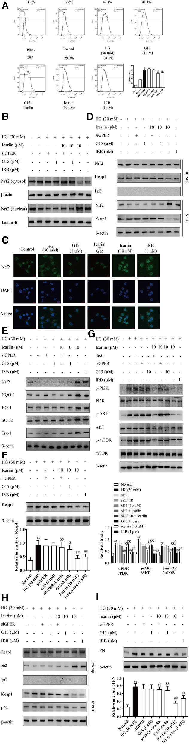
Effect of icariin on GPER mediated p62-dependent Keap1 degradation and Nrf2 activation. The cells were treated with NG (5.6 mM), HG (30 mM), HG (30 mM) + G15 (1 μM), HG (30 mM) + siGPER, HG (30 mM) + icariin (10 μM) + G15 (1 μM), HG (30 mM) + icariin (10 μM) + siGPER, HG (30 mM) + icariin (10 μM) and HG (30 mM) +IRB (1 μM) for 48 h. (A) The ROS was detected by flow cytometry. (B) the nuclear expressions of Nrf2 induced by high glucose in HMC by western blot. (C) Distribution of Nrf2 in HMC through Immunofluorescence (D) the interaction between Nrf2 and Keap1 induced by high glucose in HMC. (E) The expression of Nrf2 and antioxidant enzymes HO-1, NQO1, SOD2 and Trx1 in HMC. (F) Keap1 protein level induced by high glucose in HMC. (G) Effect of icariin on phosphorylated and total PI3K, Akt, and mTOR1 protein level induced by high glucose in HMC. (H) The interaction between Nrf2 and p62 induced by high glucose in HMC. (I) Expression of FN protein expression by western blot. Data are expressed as mean ± SD. **p < 0.01, compared with the normal control group; #p < 0.05, ##p < 0.01, compared with the Model group or HG group; $p < 0.05, $$p < 0.01, compared with the icariin group.