
FIGURE 4.
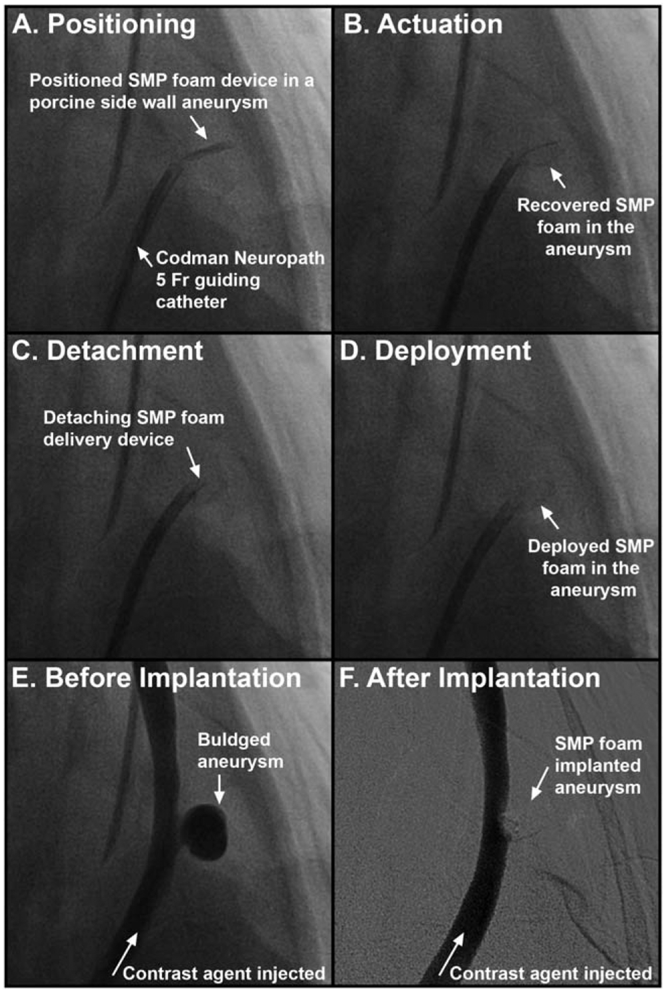
Fluoroscopic imaging of the SMP foam treatment process. The SMP foam is positioned via guide catheter in the aneurysm sac (a). As the SMP foam recovers or actuates to its expanded shape (b), the foam friction decreases allowing detachment from the delivery device (c) and release in to the aneurysm (d). Contrast dye injections are used to visualize the aneurysm before (e) and after (f) treatment with SMP foam.