
FIGURE 5.
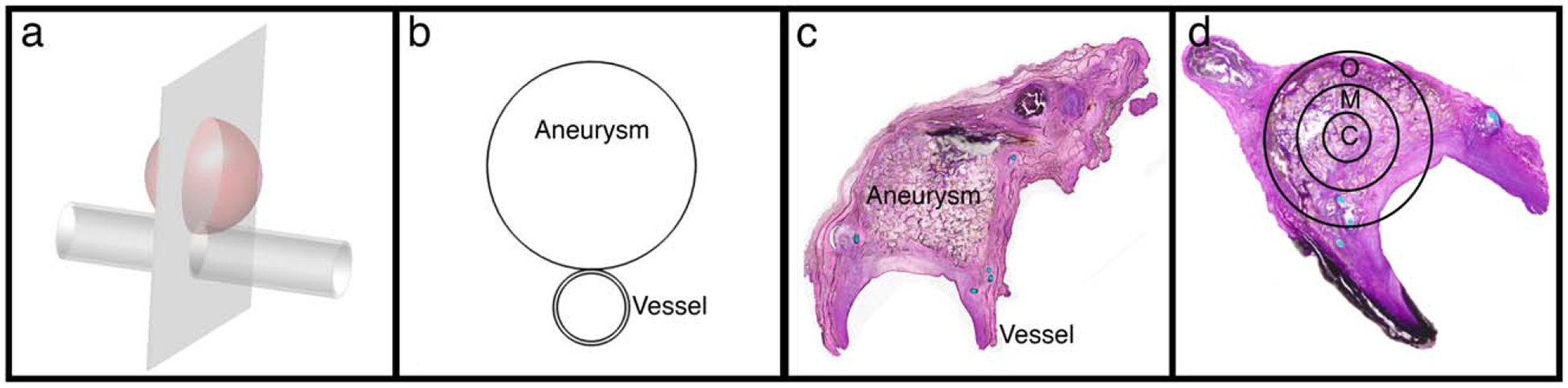
Histology sectioning and evaluation. (a) Schematic of vessel with aneurysm showing plane of histology sections; (b) Resulting cross-section of aneurysm and vessel for histology slides; (c) Example histology section; (d) Definition of zones used for evaluating each histology section: C – Central, M – Mid, O – Outer.