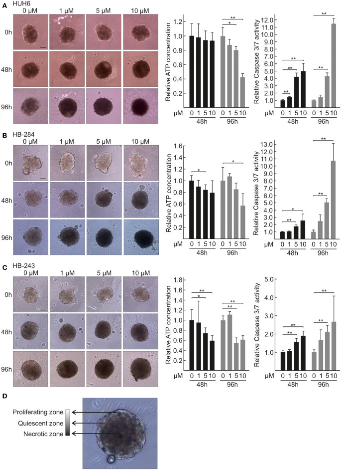
Figure 1.
Morphology, viability, and caspase 3/7 activation of HB spheroids treated with CQ. Morphology of HUH6 (A), HB-284 (B), and HB-243 (C) derived spheroids treated with control medium (0 μM) or CQ at concentrations of 1, 5, and 10 μM (left panel). Relative ATP concentration (middle panel) and relative caspase 3/7 activation (right panel) in HUH6 (A), HB-284 (B), HB-243 (C) derived spheroids after 48 and 96 h CQ treatment. CQ concentrations; 1, 5, and 10 μM. *P < 0.05. **P < 0.01. Statistical significance was assessed with one-way ANOVA. Bar plots are presented as relative values of mean ± RSD (N = 3). Characteristics of proliferating, quiescent, and necrotic spheroid morphology (D). Pictures were captured at initiation of treatment (0 h) and after 48 h and 96 h of CQ administration. Magnification 10×, scale bar = 10 μm.

