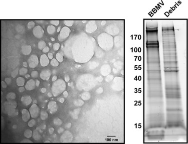
Figure 1.

Preparation and analysis of T. ni midgut brush border membrane vesicles (BBMV). (A) Transmission electron microscopic image of negatively stained BBMVs. (B) SDS‐PAGE gel of proteins from 4th‐instar BBMV or cell debris remaining after BBMV isolation. The gel shows enrichment of proteins in the BBMV fraction as reflected by different protein banding patterns in the BBMV and remnant midgut cell debris preparations. The size (kDa) of molecular weight markers is shown in the left hand margin.
