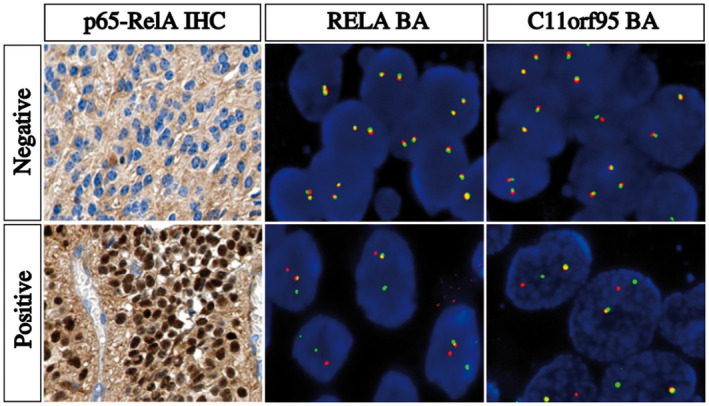
Figure 2.

Detection of NFκB pathway activation by IHC and RELA and C11orf95 rearrangements by FISH. IHC and FISH images showing a negative case (top panel) and a positive case (bottom panel). Left panel, p65‐RelA IHC images showing an intense nuclear staining in a positive case reflecting an activation of NFκB pathway and a negative case without nuclear staining. Middle panel, representative image of a slide hybridized with a RELA Break‐Apart FISH probe. In this given example, the images show nuclei harboring a split (red and green signals) and a fused signal in a positive case and two intact fused signals in a negative case. Right panel, representative image of a slide hybridized with a C11orf95 Break‐Apart FISH probe. In this given example, the images show nuclei harboring a split (red and green signals) and a fused signal in a positive case and two intact fused signals in a negative case. IHC, original magnification x40. FISH, Original magnification x1000. IHC, immunohistochemistry; FISH, fluorescence in situ hybridization
