Figure 3.

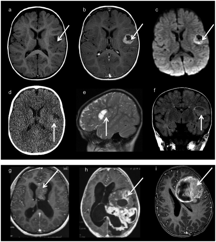
Neuroimaging findings (a–f): Images from one typical supratentorial RELA‐fused ependymoma. Axial T1‐weighted images before (a) and after (b) contrast material injection, axial diffusion weighted images (c), CT scan (d), sagittal T2 weighted images (e) and coronal FLAIR images (f). Cortical based, well‐demarcated solid and cystic lesion with a mural nodule and minimal peripheral edema. Contrast injection enhances the nodule and the periphery of the cystic portion. There is diffusion restriction on diffusion‐weighted imaging (c) and a hyper density on the CT scan corresponding to the hyper cellularity. (g): Axial T1 weighted images with contrast injection corresponding to a tumor with mixed ependymal/ subependymal histological features. The intraventricular mass is solid with heterogeneous contrast enhancement. (h): Axial T1‐weighted images with contrast injection from a YAP‐fused ependymoma showing a voluminous lesion with prominent solid component with heterogeneous and multinodular appearance. (i): Axial T1‐weighted images with contrast injection from a “HGNET, MN1” tumor, showing a large lesion with a prominent solid portion and necrotic areas.
