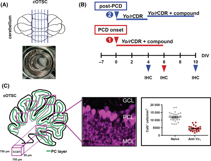
Figure 1.

Ex vivo paraneoplastic cerebellar degeneration (PCD) model. (A) The illustration shows the cutting planes of the cerebellar vermis including the incubation chamber to maintain the rat cerebellar organotypic slice culture (cOTSC), the basis of the ex vivo PCD model system. (B) Experimental design: 7 days’ postpreparation, anti‐Yo/rCDR and compounds were co‐applied either immediately at PCD onset [(1), red arrow] or 4 days after PCD induction [(2), post‐PCD, blue arrow] for 6 days. The pathological progression of anti‐Yo/rCDR internalization was determined by immunohistochemical (IHC) staining of Purkinje neurons with biomarker calbindin at 4, 6 and 10 days of treatment. (C) Three to eight confocal images of 750 × 750 × 50 μm in each cOTSC slice for each treatment were scanned and the Yo antibody incorporation‐dependent slice pathology (4, 6 and 10 days) was analysed by counting Purkinje neurone somata in the Purkinje cell layer (PCL) immunohistochemically visualized by biomarker calbindin D28K (magenta). Calbindin‐positive cells (Calb+) were calculated as number of Calb+ cells/mm3 and plotted for each group.
