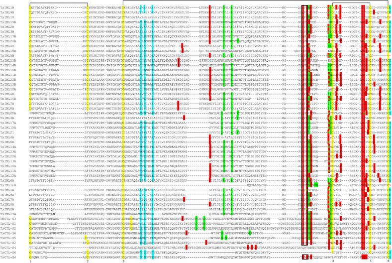
Figure 4.

Multiple sequence alignments of C‐type lectin domains (CTLDs) in Thitarodes xiaojinensis. The conserved cysteine (Cys) residues are highlighted in yellow and marked with 1 to 6. The predicted disulfide linkages between Cys‐1 and ‐2, ‐3 and ‐6, ‐4 and ‐5 are shown by lines. The motifs (e.g. EPN and QPD) that usually participate in carbohydrate recognition in CTLDPs are indicated by the enclosed box. Residues involving in binding Ca2+ in site‐1 and site‐4 are shaded in green and cyan, respectively. Ligand binding sites in each CTLD were predicted through COACH combining with I‐TASSER server. The consensus binding residues are shaded in red, with those also ligating to Ca2+ in bold and green font.
