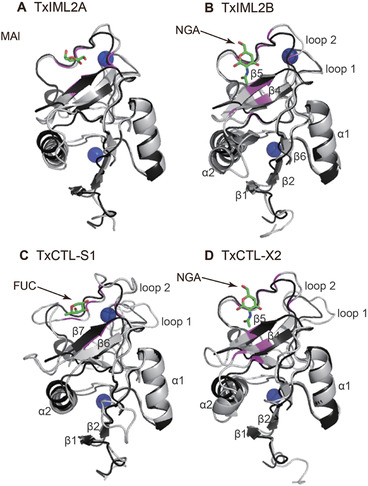
Figure 5.

Structures of the C‐type lectin domains (CTLDs) in Thitarodes xiaojinensis immulectin (IML)2A (A), IML2B (B), CTL‐S1 (C) and CTL‐X2 (D) and structural comparison with the CTLD in human human dendritic cell‐specific intercellular adhesion molecule‐grabbing nonintegrin‐related (DC‐SIGNR) (1K9J). T. xiaojinensis and human CTLDs are colored gray and black, respectively. Secondary structure elements in Figure S1 are labeled in the corresponding models. The blue spheres indicate Ca2+ ions. The stick models represent the carbohydrate ligands (the C atoms are shaded in green and the O atoms are in pink). The residues involving in binding carbohydrate (Table S5) are highlighted in magenta. MAN, alpha‐D‐mannose; NGA, N‐acetyl‐D‐glucosamine; FUC, alpha‐L‐fucopyranose.
