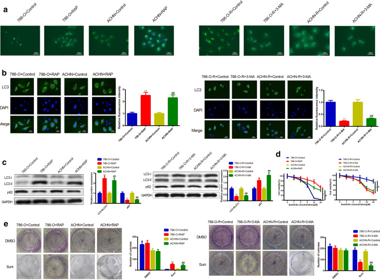
Fig. 3.
Effect of cell autophagy on sunitinib resistance in RC cells. To verify the regulatory role of cell autophagy in sunitinib resistance of RC cells, the parental cells (786-O and ACHN) were subjected to RAP for activation of cell autophagy. Meanwhile, sunitinib-resistant cells (786-O-R and ACHN-R) were exposed to 3-MA for inhibition of cell autophagy. Increased autophagic vacuoles of parental cells after RAP treatment as well as reduced autophagic vacuoles of sunitinib-resistant cells after 3-MA subjection were conducted by MDC staining (a). The enhanced fluorescence intensity of LC3 in RAP-induced group and suppressed fluorescence intensity of LC3 in the 3-MA-treated group were estimated by immunofluorescence (b). Protein expressions of LC3-II/LC3-I and p62 were assessed by Western blot. RAP potentiated LC3-II/LC3-I protein levels and lowered p62 expression in 786-O and ACHN cells. In 786-O-R and ACHN-R cells, 3-MA elevated protein expression of p62 and restrained LC3-II/LC3-I levels (c). CCK-8 assay was utilized to test the RC cell sensitivity to sunitinib. RAP was found to enhance the sunitinib resistance of parental cells, while 3-MA was discovered to potentiate the sunitinib sensitivity of sunitinib-resistant cells (d). Colony formation was applied to observe the formation of clone after exposure to 10 μM of sunitinib or DMSO. RAP-treated group had increased colony formation, while 3-MA-treated group possessed lowered colony formation (e). *P < 0.05, **P < 0.01, compared to 786-O + Control group or 786-O-R + Control group; #P < 0.05, ##P < 0.01, compared to ACHN + Control or ACHN-R + Control group; RC, renal cancer; RAP, rapamycin; 3-MA, 3-methyladenine

