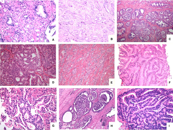
Figure 2.

Depiction of grade 4 subpatterns: (A) haphazardly distributed poorly formed very small‐sized distinct glands showing some lumen‐formation, (B) fused small‐sized lumen‐containing glands in a retiform pattern, (C) focus of glomeruloid structures within small‐ to medium‐sized distinct glands, (D) expansile rounded tumour area with cribriform pattern lacking intervening stroma or capillaries, (E) abortive glands consisting of structures with glandular shape, but lacking a lumen, to be distinguished from solid pattern grade 5 carcinoma, (F) large‐sized glands which merge together, constituting the large‐fused pattern, (G) complex fused glands with irregular cribriform area, but with intervening stroma and capillaries, (H) large‐sized glands with glomeruloid features showing a cribriform pattern, adjacent to small‐sized glomeruloid glands and (I) papillary pattern lined by columnar tumour cells reminiscent of ductal adenocarcinoma.
