
Figure 4.
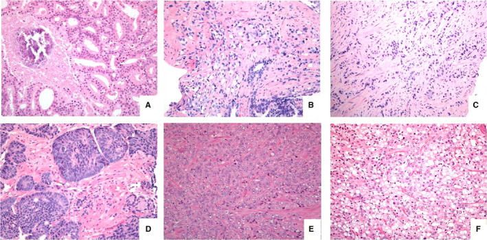
Most common grade 5 patterns, such as (A) comedocarcinoma, showing a necrotic central plug surrounded by fused glands, (B) single cells with vacuolisation with haphazard distribution between benign glands, (C) single file of tumour cells, lacking lumina, embedded in dense stroma, (D) smaller solid areas (upper right) with some acinar (pseudo‐rosetting) nuclear arrangement, but lacking lumen formation, (E) larger sheet of poorly differentiated tumour cells and (F) solid area of cells with signet ring‐like appearance.