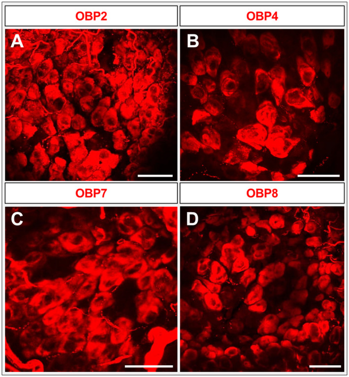
Figure 7.

Expression of odorant‐binding proteins (OBPs) from different subfamilies. (A–D) Whole‐mount fluorescence in situ hybridization utilizing maxillary (A, B, D) and labial (C) palps of the desert locust using digoxigenin‐labelled riboprobes of subfamily I‐B and subfamily III OBPs (2, 4, 7 and 8). OBPs are visualized by red fluorescence. All four OBPs are expressed in a higher number of cells, similar to OBP6 in Fig. 4. Images represent projections of confocal image stacks representing different optical layers. Scale bars: A–D, 50 µm.
